 CONFERENCE
Project title:
"Time-Resolved Autofluorescence Methodology for Non-Invasive Diagnosis of Skin Cancer."
Project contract number:
1.1.1.2/VIAA/1/16/014
Project partners:
Vytautas Magnus University, Department of Biology, Department of Biology (Vytautas Magnus University), Kaunas, Lithuania.
International Laser Center, Bratislava, Slovakia.
University of Gothenburg, Center for Skin Research, Department of Chemistry and Molecular Biology (SkinResQU, Department of Chemistry & Molecular Biology, University of Gothenburg), Gothenburg, Sweden.
Project implementation deadline:
01.01.2018 – 31.12.2020
Total project funding:
133 805,88 EUR
ERAF funding: 113 734,99 EUR
State budget financing: 13 380,58 EUR
UL funding: 6 690,31 EUR
Research manager:
Aleksejs Ļihačovs (Biophotonics laboratory)
About the project:
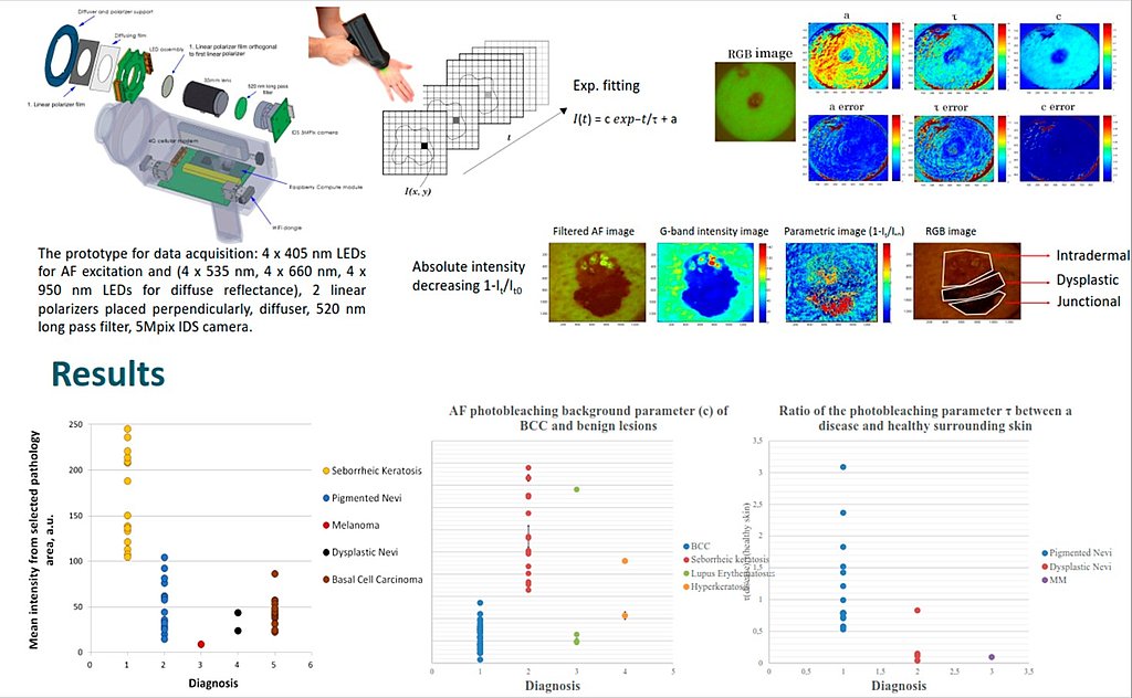
The project aims to develop a cost-effective, innovative, non-invasive methodology for the early diagnosis and control of skin cancer. The diagnostic process will consist of quantitative and qualitative assessment of biochemical and physiological function of tissues by a combination of laser-induced autofluorescence (AF) degradation and AF photo-fading techniques.
Project results:
2 original scientific articles with a high citation index, 4 conference abstracts, 1 popular science article, 3 mobility trips and scientific networking.
Acknowledgement for publications:
This work has been supported by European Regional Development Fund project “Time-resolved autofluorescence methodology for non-invasive skin cancer diagnostics” (No. 1.1.1.2/16/I/001, agreement No. 1.1.1.2/VIAA/1/16/014).
1st quarter (01.01.2018 - 31.03.2018)
Project launch and adaptation of the work plan to the new project start date. Preparation of amendments to the contract. The first month of the project was started with literature research on a topic related to the project, namely - fluorescence photobleaching mechanisms, generation of reactive oxygen species under continuous optical radiation, autofluorescence kinetics under continuous excitation, skin cancer optical diagnostic methods and data processing and others. Work has been started with the existing TCSPC (time correlated single photon counting) equipment of the IAPS UL, specifically work has been started with the aim to improve the existing method for simultaneous registration of skin fluorescence lifetime and fading parameters. The impact on the fading parameters from the fluorescence decay distribution. Measurements have been performed to find optimal parameters for recording in-vivo skin autofluorescence lifetime and photobleaching parameters. The ability of the TCSPC device to perform repeated measurements with a high repetition frequency (> 10E-3-7 sec.) is tested. In addition, the device is tested using narrowband filters, broadband filters and a monochromator. Work was started on the development of data processing scripts in order to process the results and obtain the following parameters - fluorescence lifetime, photobleaching parameters and changes in fluorescence intensity shortly after excitation.
2nd quarter (01.04.2018 - 30.06.2018)
During the reporting period, the methodology was improved for the simultaneous registration of skin laser-induced autofluorescence lifetime and photobleaching parameters. Existing data processing algorithms were improved, in particular the timing of registration was improved. Innovative data processing algorithms were proposed, which allow to view the parameters of the excited autoflurescence (lifetime of the excited state, amplitudes, photobleaching rate) depending on the time gate. This approach allows the recording of independent dynamic processes, such as the autofluorescence parameters of different natural fluorophores. It has been found that in in-vivo experiments on human skin, the kinetics of autofluorescence photobleaching at different time gates are different, which indirectly characterizes the concentrations of natural fluorophores and photochemical processes in the skin. On the other hand, in cases when the kinetics are the same, the resulting kinetics is well described by mono exponential recovery. This fact may indirectly indicate the number of natural fluorophores, or different photochemical processes. In addition, photo-bleaching experiments were performed using post-mortal pig skin. It has been found that after the laser is switched off, the intensity of autofluorescence does not recover, but continues to decrease. This result, in turn, indicates a chemical process that continues even after the laser is turned off. During this period, a mobility trip to Vytautas Magnus University, Department of Biology was implemented to implement the project task T1.2. During the mobility, as a result of several discussions (networking), an experimental design was developed to determine the possible correlation between autofluorescence photobleaching parameters and the identification of free radicals at the cellular level. The host demonstrated an experiment with the presence of exogenous sensitizers and the resulting photochemical reaction with subsequent singlet oxygen generation. Work began on the development of a methodology for the in-vitro determination of active forms of oxygen.
3rd quarter (01.07.2018 - 30.09.2018)
During the reporting period, the development of the methodology for the simultaneous registration of skin laser-induced autofluorescence lifetime and photobleaching parameters was continued. Another mobility trip to Vytautas Magnus University, Department of Biology was implemented to implement the project task T1.2. During the mobility, several discussions led to the development of an experimental design, the selection of the necessary reagents, cell cultures, excitation and detection wavelengths. A measurement strategy was developed to correctly detect autofluorescence parameters from cells and fluorescence emission parameters from SOSG sensor. As a result, the conference thesis was submitted: Alexey Lihachev, Mindaugas Tamošiūnas, Saulius Šatkauskas, Vanesa Lukinsone, and Ilze Lihacova. Generation of reactive oxygen species during autofluorescence photobleaching in-vitro. Int. Conf. Laser Applications in Life Sciences (LALS) 2018. Work was continued on the preparation of the device for fluorescence lifetime and photobleaching measurements. In particular, optical filters and excitation wavelengths corresponding to the absorption band and the fluorescence emission spectrum of the SOSG sensor were selected. Thesis published in the conference materials of “Northern Optics 2018”: Janis Spigulis, Uldis Rubins, Edgars Kviesis-Kipge, Andris Grabovskis, Aleksejs Lihacovs, Ilze Lihacova, Dmitrijs Bliznuks, Ilona Kuzmina, Ilze Oshina, Marta Lange. Advanced imaging technologies for remote assessment of in-vivo skin. Northern Optics & Photonics 2018, 12-14 September 2018, Conf. Proc., P. 94. Available at:
photonicsweden.org/wp-ontent/uploads/NorthernOptics2018_proceedings.pdf
4th quarter (01.10.2018 - 31.12.2018)
Parallel measurements of fluorescence photobleaching and decay kinetics have been performed using lasers of different wavelengths for fluorescence excitation. In the period from 08-19.10, a business trip to the University of Oulu took place with the aim to visit the Optoelectronics and measurement techniques laboratory, led by Igor Meglinski. Networking was carried out with scientists from the University of Oulu. During the mission, the laboratory was visited, the workbenches of various experiments and the optical process simulation platform (http://www.biophotonics.fi/) were viewed. During the scientific networking, we discussed research trends and priorities in biophotonics. We define synergistic research areas with the aim of attracting external funding in the future by jointly participating in European project competitions. Simulation tests of optical processes (simulation of autofluorescence kinetics in skin layers) were performed using a platform developed by scientists from the University of Oulu. In cooperation with the project partners (Vytautas Magnus University) a measurement protocol (D1.1) was developed to test the project hypothesis - tissue / cell natural fluorophores act as acceptors of optical radiation, which causes the generation of active forms of oxygen in the photosensitization process. Fluorescence spectral data of cells and SOSG have been obtained. During the experiments I have encountered several problems and ambiguous results that require interpretation. In-vitro cells have been found to emit natural autofluorescence whose spectral properties, or spectra, overlapped with the fluorescence of the SOSG sensor. When analyzing steady state spectra, it is not possible to distinguish cell autofluorescence from SOSG fluorescence. To solve this problem, an experiment was prepared in which the lifetime of the fluorescence-induced state was additionally analyzed. It is known that the degradation of natural fluorescence of cells can be described by two / three processes where each process can be characterized by lifetime (~ 0.4 ns, 1.2 ns, 7 ns). In contrast, the fluorescence decay time of SOSG is expected with a longer lifetime of ~ 10 ns. However, literature studies did not reveal the life expectancy of SOSG. SOSG has also been found to be an unstable substance (photobleaching occurs upon irradiation), leading to changes in the distribution of fluorescence intensity and changes in fluorescence degradation. These are all dynamic processes that affect the life of SOSG. At present, it is necessary to perform an experiment to determine the SOSG fluorescence lifetime intervals at specific optical radiation parameters (power, dose).
5th quarter (01.01.2019 - 31.03.2019)
In January, an algorithm for skin autofluorescence kinetics data processing was tested. An empirical algorithm was proposed that processes changes in autofluorescence intensity and calculates photobleaching parameters. The algorithm was tested on clinical data obtained in collaboration with ERDF 1.1.1.1 # 197 project. Specifically, sequential images of autofluorescence of RGB skin pathologies at 405nm excitation were analyzed. The results were summarized and the conference thesis was submitted to the ECBO conference. The conference thesis will be published in the SPIE Proc thesis collections, which are indexed in the SCOPUS database.
Fluorescence emission kinetics of endogenous fluorophores were performed with an externally connected singlet oxygen sensor (SOSG). In-vitro Me45 melanoma cells were used in the study. Cell fluorescence kinetics tests were performed using a 405 nm laser excitation. During the study, it was found that the excitation of autofluorescence of cells decreases exponentially in the uninterrupted 405nm laser excitation. In contrast, the fluorescence intensity of the added SOSG sensor increases exponentially. The obtained data indicate the formation of singlet oxygen in the process of photobleaching at the cellular level. The obtained results confirm the hypothesis of the project that endogenous fluorophores transfer absorbed energy to oxygen and convert it into oxygen radicals. In addition, in-vitro measurements of singlet oxygen under optical radiation and ultrasound were performed. The conference thesis was submitted to the DOC RIGA 2019 conference on the obtained results. As a result of networking with researchers from Vytautas Magnus University, additional oxygen radical sensors were selected which will be used to more accurately identify possible processes under the influence of optical radiation.
Work continued on in-vitro data processing of autofluorescence spectra of cells. In collaboration with researchers from Vytautas Magnus University, I performed endogenous fluorophore photobleaching measurements to identify the correlation of fluorescence kinetics with cellular metabolic activity and hypoxia. In the period from 24 to 29, I networked with Bulgarian scientists. I present my postdoctoral project E.D. At the seminar of the Institute of Electronics. During the mission, I received consultations on endogenous skin fluorescence in UV / VIS excitation. Fluorescence measurements were performed using ex-vivo skin pathologies at 405 nm and 785 nm laser excitation. I process ex-vivo skin pathology fluorescence data.
6th quarter (01.04.2019 - 30.06.2019)
From 07.04 to 12.04, as part of a mobility trip, I visited the International Laser Center (Bratislava, Slovakia). During the mobility I presented my postdock project at the ILC (International Laser Center) seminar. During the mobility I am introduced to the ILC research equipment and its ability to perform spectral and time-resolved measurements and analysis of results. During the networking, the aim of the project was discussed and several hypotheses were put forward as well as experimental protocols were drawn up. During mobility, fluorescence measurements were performed with time resolution using different samples (isolated fluorescent molecules, ex-pig skin). The lifetime and photobleaching kinetics of autofluorescence were recorded, and the recovery of autofluorescence after photobleaching was also examined. The fluorescence degradation and photobleaching kinetics of fluorescent molecules under the influence of various external factors were recorded. The measurements used CLSM (confocal laser scanning microscope) and TCSPC (Time-Correlated Single Photon Counting) equipment. In the period from 15.04 to 30.04 I processed the obtained data. In the period from 01.05 to 06.05 I processed the fluorescence spectra of endogenous fluorophores. Annual leave from 07.05.2019 to 16.05.2019 and from 20.05.2019 to 02.06.2019. On May 17, I present the results of the project on the UL Technology Day. In June, I validated the photobleaching algorithm using autofluorescence images of skin pathology obtained in 405nm LED excitation. The results of the algorithm validation were summarized in a bench report. In the period from 23.06.2018 to 27.06.2019, I attended the European Biomedical Optics Conference (ECBO 2019, Munich, Germany) and presented a poster “Imaging of LED-excited autofluorescence photobleaching rates for skin diagnostics”.
7th quarter (01.07.2019 - 30.09.2019)
In July, I processed the accumulated experimental data with the aim to identify possible free ones that are generated in cells and tissues during the photo-fading process. The results and methodology of the study have been summarized, as a result of which the conference thesis “Investigation of ROS generation in cells and tissues during autofluorescence photobleaching” has been submitted for participation in the SPIE Photonics West 2020 conference. In September, I performed an analysis of the fluorescence spectra of endogenous fluorophores and their lifetimes. From 08.09-12.09 I attended the training school Multimodal Optical Imaging, Trnava, Slovakia. I continued to process and analyze the accumulated experimental data in order to find a correlation between the metabolic state of biological tissues and their autofluorescence properties. Participation with an informative poster about the activities of the postdoctoral project on Scientists' Night 27.09.2019.
8th quarter (01.10.2019 - 31.12.2019)
During the reporting period, I investigated the correlation between endogenous fluorophore AF-induced lifetime and their photobleaching parameters. Analyzing the fluorescence lifetime of endogenous fluorophores before and after photobleaching, it was found that the lifetime of the excited state decreases in the photobleaching process. The longer life component decreases relatively sharply. In several tests, it has been found that longer component lifetime changes in the photobleaching process could be used as a potential diagnostic criterion in in-vivo skin diagnostics. In parallel detection of AF lifetime and photobleaching in in vivo skin samples, it was found that the distribution of lifetime and photobleaching parameters in healthy and abnormal skin differs dramatically (Milestone 2.1). In collaboration with the International Laser Center, it has been concluded that subjecting one type of fluorophore (flavin) to in vitro tissue is characterized by an inhomogeneous decrease in fluorescence intensity. In addition, functional changes in these fluorophores are observed, while morphological changes were not observed. Due to this effect, photobleaching could potentially be used as an alternative method to assess the metabolic activity of endogenous fluorophores. From October 9-11, I visited the European Laboratory of Nonlinear Optics (LENS, Florence, Italy) and networked with scientists from the University of Florence. From 11 to 15 November networking with a scientist from the Bulgarian Academy. Experiments were performed using ex-vivo samples of skin and skin pathologies. Ex-vivo skin fluorescence spectra were obtained under UV / VIS and IR excitation. From 18 to 23 November networking with scientists from the University of Samara. Experiments using in-vitro cells were performed in collaboration with scientists from the University of Samara. Fluorescence spectra in IR excitation were obtained. In December, the methodology was improved for the simultaneous registration of in-vivo skin laser-induced autofluorescence lifetime and photobleaching parameters. Existing data processing algorithms were improved, in particular the timing of registration was improved. Innovative data processing algorithms were proposed, which allow to view the parameters of the excited autoflurescence (lifetime of the excited state, amplitudes, photobleaching rate) depending on the time gate.
9th quarter (01.01.2020 - 31.03.2020)
WP3 Ex-vivo / in-vivo study of AF lifetimes and photobleaching rates of different skin layers / In collaboration with the University of Gothenburg, a description of the planned experiment and a work plan have been prepared and agreed (attached to the outcome documents). Due to the emergency situation caused by Covid-19, the cooperation agreement between the University of Latvia and the University of Gothenburg is not being signed at the moment, as my mobility dates cannot be foreseen. Consequently, the mobility trip to the partner (University of Gothenburg) is currently being moved to the autumn, it was planned at the beginning of the 2nd quarter. It is difficult to predict whether a mobility trip will take place at all. It is hoped that the situation will improve significantly after the summer.
WP4 Development of Data Processing Algorithms and Clinical Validation of Methodology / During the reporting period two conference theses were prepared and submitted: 1) “Non-invasive multimodal imaging system for skin cancer detection,” The 2nd World Congress on Advanced Treatments in Skin Cancer (Skin-Cancer2020, Berlin, Germany, April 2-3). Due to Covid-19, the Congress was canceled indefinitely, but the abstract was adopted, it will be presented as soon as possible. 2) “Optical multimodal non-invasive diagnostics of skin cancer,” International Conference on Biomadical Photonics 2020, (Montpellier, France, 16-18 April). The abstract was adopted, but due to Covid-19, the conference was canceled and carried over to the following year. A popular scientific article “Portable device for early noninvasive diagnosis of skin cancer” has been published in the Yearbook of the Latvian Academy of Sciences 2020, pp. 56-61, (http://www.lza.lv/images/stories/YearBook_2020.pdf). Participation in the 78th LU conference in 2020. On February 14, the 2nd annual report of the project was presented with a poster presentation on the results of the project, as well as for the evaluation of the Scientific Council. Clinical measurements were performed at the Latvian Oncology Center (RAKUS) using a prototype for obtaining autofluorescence photobleaching images in 405 @ LED excitation. Possibilities to process and segment the obtained images using artificial neural networks and a machine learning approach were considered. During the reporting period, a collaboration was established with the Dermatooncology Clinic of Semmelweis University (Md. Norbert Kiss, Budapest, Hungary). Clinical imaging is currently underway in Hungary using a cloud based portable device for skin multispectral and autofluorescence imaging. In collaboration with Semmelweis University, several options for using photobleaching analysis for rare disease screening are being considered. The possibility of differentiating cutaneous angiokeratomas from other vascular diseases is being investigated with a view to early detection of Fabry disease. Clinical measurement images are taken in Latvia and Hungary and stored and processed using the cloud based platform https://checkyourskin.eu/ (Dermation Kinetics Data Set D4.2 of Skin Formations).
10th quarter (01.03.2020 - 30.06.2020)
Leave from 1.04.2020 to 30.04.2020. For clinical validation of data processing algorithms, autofluorescence images accumulated at the Latvian Oncology Center in Riga were used, which were taken with a prototype created in synergy with the ERDF project “Portable device for early contactless diagnosis of skin cancer”. At the moment, data collection at the Latvian Oncology Center does not take place due to the consequences of covid-19. Thanks to the collaboration with the Faculty of Dermatology, Venereology and Dermatooncology, the acquisition of clinical data is being continued at the University of Semmelweiss, Budapest, Hungary, using a prototype for skin autofluorescence imaging and a macrocomputing service (https://checkyourskin.eu/). For automatic calculation of autofluorescence photobleaching parameters for skin cancer diagnosis and classification, a skin and formation segmentation algorithm is developed using artificial neural networks. Three theses were submitted for the conference Biophotonics Riga 2020, which will take place on 24-25. August, Riga, Latvia: A. Lihachev, I. Lihacova, M. Lange, E. V. Plorina, E. Cibulska, D. Bliznuks, A. Derjabo, N. Kiss, “Imaging of LED excited autofluorescence for skin lesions assessment.” P. I. Lihacova, A. Lihachev, M. Lange, E. V. Plorina, E. Cibulska, D. Bliznuks, A. Derjabo, N. Kiss, “Optical multimodal non-invasive diagnostics of skin cancer.” P. M. Lange, S. Bozsányi, E.V.Plorina, A.Lihacovs, A. Derjabo, “Spectral imaging as a tool for the evaluation of skin cancer post-operative scars”. P.